

Treatment
Pediatric Nephrostomy Tube
One major function of the kidney is to filter blood and produce urine. Urine flows from the kidneys to the urinary bladder via a small tube called the ureter. Sometimes the normal flow of urine becomes obstructed due to stones, congenital abnormalities, abdominal and pelvis masses, or trauma. This obstruction to the flow of urine can lead to infection and loss of kidney function.
By utilizing a multidisciplinary approach in conjunction with experts in Interventional Radiology, Urology and Nephrology at Children's National Hospital, we formulate a treatment plan to preserve kidney function. This plan may require percutaneous nephrostomy tube placement, a small tube placed through the skin of the lower back into the kidney under ultrasound and fluoroscopic guidance. Urine will then drain into a small bag external to the patient. A nephrostomy tube may be in place for days, weeks or months. The decision to remove the nephrostomy tube is also made after consulting with these teams.
Frequently Asked Questions
What is a nephroureteral stent in children?
What is a suprapubic cystostomy tube in children?
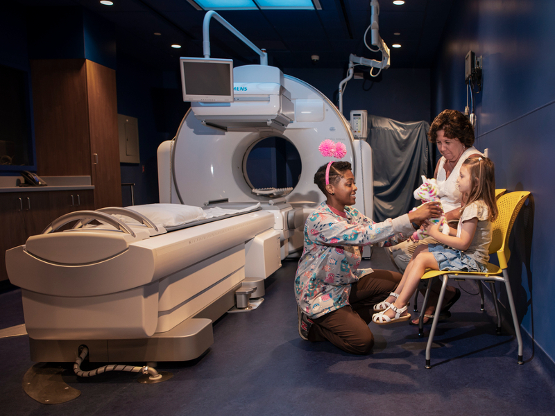
How is a nephrostomy tube insertion performed in children?
Will my child be awake during the nephrostomy tube procedure?
How long will my child's nephrostomy tube procedure take?
Will my child be in any pain after the nephrostomy tube procedure?
What are the risks of a nephrostomy tube in children?
What happens after my child's nephrostomy tube procedure?
When can I remove my child's bandages after a nephrostomy tube procedure?
When can my child bathe after a nephrostomy tube procedure?
How and when will my child's nephrostomy tube be removed?
Are there any activity restrictions after a nephrostomy tube procedure in children?
Meet the Providers Who Offer Nephrostomy Tube
Departments that Offer Nephrostomy Tube
Interventional Radiology
Children's National interventional radiologists perform a full range of minimally invasive, image-guided procedures to both diagnose and treat disease in infants, children and adolescents. Learn more about how we help children in our care.

Help Kids and Make a Difference
Invest in future cures to help children have brighter futures.