

Treatment
Pediatric Gastrojejunostomy Tube Placement (GJ Tube)
A gastrojejunostomy tube placement (GJ tube) is just like a G-tube except that it contains an extra portion of tubing that extends from the stomach to the intestine. An extra side port allows feeding and medication delivery directly into the intestine through extension tubing. Your family will receive education on the skin care surrounding the tube site as well as on the proper usage and maintenance of the tube and extension material prior to the procedure. You will receive more information about the type of feeds that go through G and GJ tubes. Your child will often see pediatric experts from Clinical Nutrition Services at Children's National Hospital after a G or GJ tube placement.
If your child requires a GJ tube, typically a G-tube will be placed first, and then after six to eight weeks the tube can be converted from a G-tube to a GJ tube. However, on occasion a direct GJ tube will be placed. The Interventional Radiology team at Children's National Hospital will begin planning the approach by doing an ultrasound study of your child’s abdomen, which will help us identify the track to the stomach and the location of vital structures (such as other organs and blood vessels) to be avoided during the procedure. Then, we place a small tube through the nose into the stomach and fill the stomach with air. This makes the stomach an easier, more visible target for puncture with a needle from the skin. Once we have marked a spot for the feeding tube, we will inject a local numbing medicine into the skin. Your child's interventional radiologist will use live X-ray (fluoroscopy) to place a needle through the skin into the stomach. After the needle is in the stomach, the GJ tube will be inserted over a guide wire into the intestine. The doctor will inject contrast during X-rays to ensure the end of the tube is in the intestine.
Frequently Asked Questions
How is a G-tube converted to a GJ tube for children?
Will my child be awake during the GJ tube procedure?
Will my child be in any pain during the GJ tube conversion?
How long does a GJ tube conversion in children take?
What are the risks of GJ tube conversion in children?
Meet the Gastrojejunostomy Tube Placement (GJ Tube) Providers
Departments that Offer G-J Tube
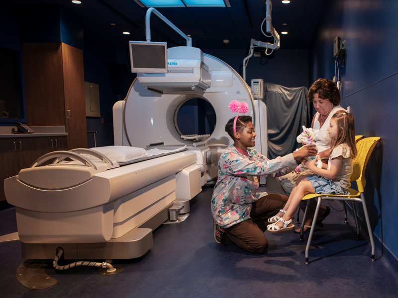
Interventional Radiology
Children's National interventional radiologists perform a full range of minimally invasive, image-guided procedures to both diagnose and treat disease in infants, children and adolescents. Learn more about how we help children in our care.

Help Kids and Make a Difference
Invest in future cures to help children have brighter futures.