Treatment
Cardiac First Trimester Imaging
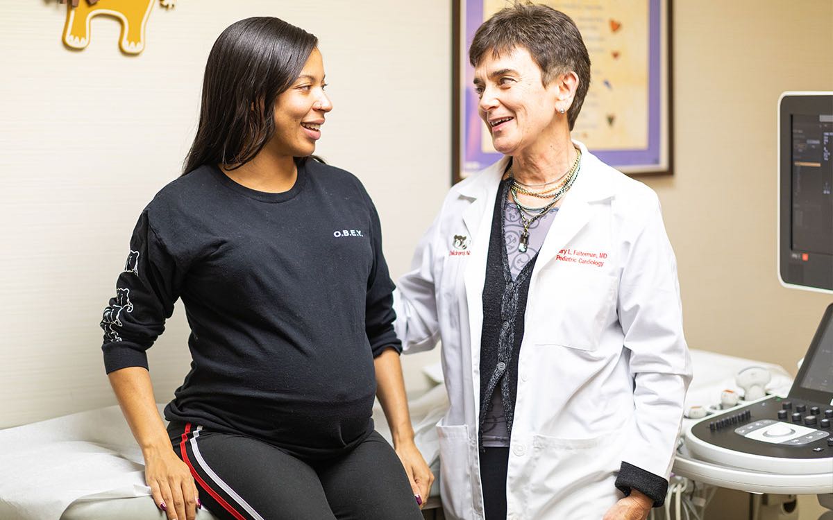
The Prenatal Cardiology Program at Children’s National Hospital offers innovative and state-of-the-art care. With advancing technologies, imaging of the fetus has improved substantially in the past 5 years. With new ultrasound equipment that has very high resolution and techniques such as transvaginal ultrasound, the heart may be imaged as early as 11-12 weeks gestation. The pictures that can be obtained are limited, but in most cases it is anticipated that major heart abnormalities will be identified. This service is used as an adjunct to the standard fetal echocardiogram that is done at 18-20 weeks, though seeing the heart at this early stage may be extremely helpful to select families with an understanding that the diagnosis will need to be confirmed later in the pregnancy.
- Indications for first trimester fetal echo
- Increased nuchal translucency noted in the first trimester
- Significant family history of CHD particularly complex heart disease in a previous child
- Maternal lupus, particularly if a previous child developed complete heart block
Meet the Providers Who Perform First Trimester Imaging
Departments that Offer Cardiac First Trimester Imaging

Heart and Lung Center
Our expert pediatric heart team, including more than 40 subspecialties, offer advanced heart care and excellent outcomes for thousands of children every year.

Help Kids and Make a Difference
Invest in future cures for some of life's most devastating diseases. Give today to help more children grow up stronger.


